Biography - Speakers for FluoroFest 2016
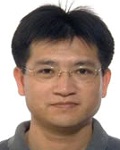
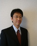
Prof. David Birch - Department of Physics, University of Strathclyde, Glasgow, UK
Djs.birch@strath.ac.uk
 David Birch is Professor of Photophysics at the University of Strathclyde.
He has published over 200 research papers, mainly on fluorescence lifetime spectroscopy. His present research focuses on fluorescence at the biomedical interface in the study of nanoparticles, melanin structure and photophysics, fibril formation in neurology, biomarkers for cancer detection and continuous glucose monitoring.
David studied physics at the University of Manchester where his PhD on diphenylpolyene fluorescence was supervised by John B Birks. After lecturing at Manchester he moved into industry to work on high-resolution organic mass spectrometry with VG Micromass Ltd. His interests subsequently returned to fluorescence when he moved to Strathclyde University where he was appointed Professor of Photophysics in 1993. David was Head of Department from 2004 -10, a period leading up to the Government-led REF 2014 research assessment in which the Department was ranked 1st for research quality across all UK university physics departments.
External appointments include: in 1999 the Sir C V Raman Endowment Visiting Chair at the University of Madras, in 2000 a Visiting Professorship at Kyoto Institute of Technology, from 2002 the Visiting Chair of Applied Physics at the Czech Technical University and in 2014 the Green Honors Chair at Texas Christian University. He is a Fellow of Scotland's National Academy and a member of the Permanent Steering Committee of the Methods and Applications in Fluorescence conference series. He is Editor-in-Chief of the Institute of Physics journal Measurement Science and Technology and joint founding Editor-in-Chief of one of the Institute's newest journals, Methods and Applications in Fluorescence. He has served on the Editorial Board of SPIE's Journal of Biomedical Optics since its launch in 1996.
David is a Director and co-founder of IBH, one of the earliest Scottish University spin-out companies. The Company manufactures fluorescence lifetime instruments and joined Horiba Scientific in 2003.
Title: Using fluorescence for nanoparticle metrology with 0.1 nm resolution
There is growing awareness of the environmental and health consequences that might be associated with the proliferation of nanoparticles, particularly those in the 1-10 nm range that can traverse cellular membranes. Hand-in-hand with this interest goes the search for improved methods of measuring nanoparticle size for research, the need for low cost, more portable and easy-to-use methods that would facilitate wider monitoring, and the need for internationally agreed standards. A recent European Commission report [1] on measurement methods for nanoparticles serves to underline the importance of the topic and provides a useful summary of the main techniques in current use such as light scattering, small angle x-ray scattering (SAX), small angle neutron scattering (SANS) and scanning and transmission electron microscopy (SEM and TEM respectively).Although fluorescence is a phenomenon that enables many different types of measurement [2], it is not usually associated with metrology. Nevertheless some years ago the Photophysics group at Strathclyde University introduced an alternative approach to conventional methods for measuring 1-10 nm nanoparticles dispersed in solution that is based on measuring the decay of fluorescence anisotropy of dye bound to a nanoparticle undergoing Brownian rotation. It has been shown to be capable of ~ 0.1 nm resolution and yet is of lower cost and easier to use than SAX, SANS, SEM and TEM. So far this has proved quite successful using a range of both electrostatically and covalently attached dyes in studies of silica nanoparticles growing during the sol-gel process [3,4] and stable LUDOX colloids that are promising metrology standards [5].
In this talk I will describe the basics behind fluorescence nanoparticle metrology, outline the main errors to be overcome and describe how recent developments in dye technology promise to make studies at the present day limit of measurement more routine.
References
- Linsinger T P J, Roebben G, Gilliland D, Calzolai L, Rossi F, Gibson N, Klein C 2012 Requirements on measurements for the implementation of the European Commission definition of the term "nanomaterial". European Commission Joint Research Centre Report. doi 10.22787/63490
- Birch D J S, Chen Y and Rolinski O J 2015 Fluorescence. Photonics: Scientific Foundations, Technology and Applications, Volume IV, Biological and Medical Photonics, Spectroscopy and Microscopy. Ed. D L Andrews. Wiley. Ch.1. 1-58.
- Birch D J S and Geddes C D 2000 Sol-gel particle growth studied using fluorescence anisotropy: an alternative to scattering techniques. Phys. Rev. E 62 2977-80.
- Yip P, Karolin J and Birch D J S 2012 Fluorescence anisotropy metrology of electrostatically and covalently labelled silica nanoparticles. Meas. Sci. Technol. 23 084003.
- Apperson K, Karolin J, Martin R W and Birch D J S 2009 Nanoparticle metrology standards based on the -resolved fluorescence anisotropy of silica colloids. Meas. Sci. Technol. 20 025310.
Dr. Yu Chen - University of Strathclyde
 Dr. Yu Chen received her PhD from Birmingham University in nanoscale physics in 2000 and subsequently was appointed to a lectureship in molecular nanometrology at the University of Strathclyde in 2007 funded by an EPSRC Science and Innovation Award. She is a fellow of the Royal Microscopical Society, a member of the European Microscopy Society, the Institute of Physics, the American Chemical Society, and the Nanotechnology Knowledge Transfer Network. She serves as an expert panel member for the European Cooperation in Science and Technology, a referee for top international journals, an editor of Sample of Science, an invited speaker, chair and organizer of a number of international conferences and workshops.
Dr. Chen's main research activities lie in the creation and characterization of nanoscale structures for their unique physical and chemical properties utilizing both optical and electron microscopy and spectroscopy. Her recent research focuses on fluorescent gold nanoparticles and surface plasmon enhanced effect, including two-photon luminescence, energy transfer and SERRS from noble metal nanoparticles, arrays and porous media, with strong link to biomedical imaging and sensing, as well as nanoparticle-cell interaction and cytotoxicity.
Title: New opportunities in biomedical imaging and sensing
Intrinsic luminescence from gold nanoparticles has attracted intensive interest in recent years. It combines with high photostability, low toxicity, tunable absorption band and ability to conjugate to bio-molecules, making gold nanoparticles a versatile probe in biological imaging and sensing. We have studied the two-photon luminescence (TPL) from gold nanorods (GNRs) and found that their characteristic short lifetime (less than 100ps) can be used to distinguish gold nanorods from other fluorescent labels and endogenous fluorophores in lifetime imaging. In addition, we have observed surface plasmon enhanced energy transfer between biological labeling dyes and gold nanorods under two-photon excitation in both solution and intracellular phases. These studies demonstrated that gold nanoparticle-dye energy transfer combinations are appealing, not only in Fluorescence Resonance Energy Transfer (FRET) imaging, but also energy transfer-based fluorescence lifetime sensing of bio-analytes. Internalization of GNRs has been studied via FRET based fluorescence lifetime imaging using GFP labelled early endosome. Observed energy transfer between GNRs and GFP indicates the involvement of endocytosis in GNR uptake. Finally, a novel nanoprobe based on gold nanorod for nucleic acid sensing has been developed. Drastic recovery of fluorescence intensity and distinctive changes in fluorescence lifetime has been observed after hybridization of nanoprobes with targeting oligonucleotide in vitro. Detection of cancer mRNA has been demonstrated at single cell level, suggesting the potential in cancer diagnosis and prognosis.Xiaohong Fang - Chinese Academy of Sciences
 Xiaohong Fang received her Ph.D. degree in Analytical Chemistry at Peking University (China) in 1996. After one-year postdoc work in Univ. of Waterloo (Canada), she worked as a research associate in Univ. of Florida (USA) from 1998 to 2001. She was recruited to the Chinese Academy of Sciences (CAS) in the "Hundred Talents Program" in December 2001, and then became a professor of chemistry in Key Laboratory of Molecular Nanostructures and Nanotechnology at Institute of Chemistry, CAS. She is also an adjunct professor of the University of the Chinese Academy of Sciences. Since 2007, she has been appointed as a chief scientist of the National Key Basic Research Program on Nanosciences by Ministry of Sciences and Technology of China.
Her main research interest is the development of new biophysical and bioanalytical methods for the real-time protein detection and for the study of biomolecular interaction at the single molecule level. This includes: single molecule imaging and monitoring in living cells, biomolecular interaction study by single-molecule force microscopy, novel molecular probes for real-time protein recognition and nanobiosenor development. She has over 170 publications in scientific journals such as PNAS?JACS?Angew. Chem. Int. Ed.?Adv. Mater., and over 20 granted patents.
Title: Monitoring Membrane Receptor Dynamics in Living Cells by Single-Molecule Fluorescence Imaging
Single-molecule fluorescence imaging of receptor dynamics is becoming a powerful tool for the real-time study of receptor activation and internalization to achieve a better understanding of signal transduction mechanism. With single-molecule imaging of green fluorescent protein (GFP) tagged transforming growth factor ß (TGFß) receptors at the cell surface, we have proposed a new model in which the activation of serine-threonine kinase receptors is accomplished via monomer dimerization. We further determined the mobility and dimerization kinetics of TßRII on cell membrane by tracking individual receptors that were site-specifically labeled with an organic dye. In addition, after monitoring caveolae- and clathrin-mediated TßRI receptor endocytosis in living cells, we uncovered the direct fusion of clathrin- and caveolae-mediated endocytic pathways during TGF-ß receptor endocytic trafficking, which could generate a new multifuntional sorting site for signal regulation.Michael GONSIOR - University of Maryland Center for Environmental Science (UMCES)
 I am a biogeochemists with an interest in the molecular diversity of complex organic matrices in aquatic environments analyzed by modern analytical technology. Broad scale fingerprinting of complex dissolved organic matter (DOM) has enabled to shed light on the chemodiversity of this material in diverse aquatic systems.
My major focus has been the marine carbon cycle, but also freshwater and engineered systems. I have been using primarily tools to determine the optical properties (absorbance, fluorescence) and the molecular composition (ultrahigh resolution mass spectrometry) of DOM. I also have a strong interest in the reactivity of DOM and in particular its photochemical degradation in aquatic systems. We recently developed a unique photo-degradation system, which is used to determine the time-resolved fate of chromophores in DOM under strictly controlled pH conditions and results indicated a strong pH dependency, which is dependent on the origin of DOM.
Title: Photochemistry of Marine Dissolved Organic Matter
The link between detailed molecular characterization of marine dissolved organic matter analyzed by ultrahigh resolution mass spectrometry and its optical properties is only slowly emerging and this seminar will give an overview about what we know about the molecular composition of light absorbing chromophoric DOM (CDOM) in the World's Oceans with a specific focus on how semi-continuous excitation emission matrix fluorescence monitoring can be used to determine photo-degradation kinetic data. CDOM is increasing with depth in the open ocean but the origin of this material is still under debate with suggested sources coming either from terrestrially-derived polyphenols or in situ production from marine autotrophs or heterotrophs. Fluorescence has been particularly useful due to its sensitivity and Parallel Factor Analysis (PARAFAC) statical modelling has advanced the field to describe different fluorescence components that were associated with different fluorophore groups. A strong correlation of fluorescence signals with the apparent oxygen utilization (AOU) in the Pacific suggested an in situ production of fluorophores, but recent results showed that this correlation is not apparent in the Atlantic. A critical discussion about potential sources of marine CDOM will also be part of this seminar.Dr.Chengzhi HUANG - Southwest University
 Dr. Huang is a Professor of analytical chemistry and pharmaceutical analysis in Southwest University, Chongqing, China.
He graduated from Southwest Normal University in 1986 with a Bachelor degree. After working there for 4 years, he pursued his Mater degree of Analytical Chemistry in Beijing Normal University, and PhD degree of Analytical Chemistry in Peking University for. During 1998.11-1999.10, he worked as a visiting researcher at the department of Analysis and Biochemistry, Central Research Laboratory, Hitachi, Ltd. (Japan). In 2000, he worked in Dr. Johne Liu in group in University of Ottawa as a short-term postdoctoral fellowship. After that, he worked in Professor Imai group in University of Tokyo for two years as a postdoctoral fellowship, engaging the synthesis of fluorescence organic reagents for analytical biochemistry.
With the support of a series of funds such as the National Science Fund for Distinguished Young Scholars, he engaged in the researches of plasmon analytical chemistry, include the bioassembly, nano analytical chemistry, and optical analytical chemistry. He has published more than 330 papers in ACS Nano, Small, Anal Chem, Chem Commun., et al, which have been cited about 5800 times with the H-index of 40.
Title: Optical Property and Analytical Applications of Carbon Dots
Since their first discovery in 2004, fluorescent carbon dots (CDs) have found to be physicochemically and photochemically stable without photobleaching, making them become a star family in biosensing and bioimaging fields.More interestingly, some colorful CDs have been prepared with the same carbon source, which are similar to the optical properties of quantum dots. However, bare CDs are usually weakly fluorescent and hard to attach functional groups on the surface.Regardless continuous efforts in circumventing the problems,challenges still remain to engineer CDs with desirable biosensing and labeling properties, such as high quantum yield, preferable selectivity, for long-term and real-time monitoring of targets of interest.Our group focuses on the optical property and application of carbon dots. It has been found that CDs presented the excitation wavelength-dependent or independent emission. To improve the quantum yield of carbon dots, carbon dots were doped with nitrogen and germanium which have been applied to biochemical analysis and bioimaging, such as phosphate,tetracycline,mercury (II).For example, a simple method for phosphate (Pi) detection was established by developing an off-on fluorescence probe of europium-adjusted CDs, which has been successfully applied to the detection of Pi in very complicated matrixes such as artificial wetlands system. This work has been highly cited and greatly appreciated.
References
- Xu, X.; Ray, R.; Gu, Y.; Ploehn, H. J.; Gearheart, L.; Raker, K.; Scrivens, W. A. J. Am. Chem. Soc. 2004, 126, 12736-12737.
- Wu, Z. L.; Gao, M. X.; Wang, T. T.; Wan, X. Y.; Zheng, L. L.; Huang, C. Z. Nanoscale 2014, 6, 3868-3874.
- Wu, Z. L.; Zhang, P.; Gao, M. X.; Liu, C. F.; Wang, W.; Leng, F.; Huang, C. Z. J. Mater. Chem. B 2013, 1, 2868-2873.
- Wei, W.; Xu, C.; Wu, L.; Wang, J.; Ren, J.; Qu, X. Sci. Rep. 2014, 4, 3564.
- Ding, C.; Zhu, A.; Tian, Y. Acc. Chem. Res. 2014, 47, 20-30.
- Trachtenberg, J. T.; Chen, B. E.; Knott, G. W.; Feng, G.; Sanes, J. R.; Welker, E.; Svoboda, K. Nature 2002, 420, 788-794.
- Yuan, Y. H.; Li, R.; Wang, Q.; Wu, Z. L.; Wang, J.; Liu, H.; Huang, C. Z. Nanoscale 2015, 7, 16841-16847.
- Zhao, H. X.; Liu, L. Q.; Liu, Z. D.; Wang, Y.; Zhao, X. J.; Huang, C. Z. Chem. Commun. 2011, 47, 2604-2606.
- Lin, M.; Zou, H. Y.; Yang, T.; Liu, Z. X.; Liu, H.; Huang, C. Z. Nanoscale 2016, 7, DOI:10.1039/c1035nr08177g.
Yunbao JIANG - Xiamen University

Kazunari MATSUDA - Kyoto University
 Kazunari Matsuda received his B.S. and M.S. degrees in applied physics from the Nagoya University in 1993 and 1995, respectively, and completed his Ph.D. in applied physics from Nagoya in 1998. He was a postdoctoral research associate in Kanagawa Academy of Science and Technology, in 1998-2004 and also appointed the PRESTO in JST in 2002-2004. He joined the Institute of Chemical Research of Kyoto University in 2004 as an Associate Professor in 2005 and promoted to Professor in 2010. He is currently a Professor in the Institute of Advanced Energy, Kyoto University. Professor Matsuda was a recipient of the Young Scientist Award, Minister of Education, Culture, Sports, Science and Technology, Japan in 2004 and Young Scientist Award, The Physical Society of Japan, in 2007.
Professor Matsuda's main research interests are the novel optical phenomena emerging in nano-materials, especially in nano-carbon (carbon nanotubes, and graphene) and atomically thin layered system by advanced laser spectroscopy. He has published about 100 scientific journals, and 100 invited talk in conferences.
Title: Novel optical phenomena in nano-carbon and atomically thin two-dimensional materials
Stephen SIMS - University of Western Ontario
 Sims received his BSc in Zoology (University of Western Ontario, 1975) and PhD in Neuroscience (McMaster University, 1982). Postdoctoral training was carried out in Physiology at The University of Massachusetts Medical School (Worcester Massachusetts) and at Brigham and Women’s Hospital (Harvard Medical School, Boston).
Sims joined Western in 1987, supported by a Medical Research Council Scholarship followed by a Scientist award. His research focuses on ion channels and calcium in muscle and bone cells. He has held support from several agencies, including Canadian Institutes of Health Research and The Arthritis Society. Sims served the academic community as Interim Chair of Physiology, Associate Vice-Provost in the School of Graduate and Postdoctoral Studies, and as a member of the Board of Governors.
Sims is chair of the CIHR New Investigator personnel committee, and is past president of the Canadian Physiological Society. He has served on review committees for CIHR, the Alberta Heritage Foundation for Medical Research, the Canadian Arthritis Network and the Arthritis Society. He has mentored 17 graduate students, 18 postdoctoral fellows, more than 50 undergraduate research trainees and 13 visiting scientists. His trainees have won national and international awards for their research, and have progressed to independent careers in academia, education, medicine, dentistry and financial markets. Sims and colleagues pioneered the use of patch clamp and fluorescence methods to study mammalian osteoclasts. They were the first to identify and characterize ion channels in osteoclasts, and their regulation. Osteoclasts are the therapy target in diseases like rheumatoid arthritis, osteoporosis and metastatic tumors in bone.
Title: Live-cell fluorescence imaging of bone cells
Professor Vivian W.W. YAM - The University of Hong Kong
 Professor Vivian Wing-Wah YAM is currently the Philip Wong Wilson Wong Professor in Chemistry and Energy and Chair Professor at HKU.
She was elected to Member of the Chinese Academy of Sciences in 2001 at the age of 38 as the youngest member of the Academy, Foreign Associate of the US National Academy of Sciences in 2012, Foreign Member of Academia Europaea (The Academy of Europe) and Founding Member of The Academy of Sciences of Hong Kong in 2015, and Fellow of The World Academy of Sciences for the Advancement of Science in Developing Countries (TWAS) in 2006. She was the Laureate of the 2011 L'Oreal-UNESCO For Women in Science Award and recipient of a number of awards and prizes, including the 2005/06 Royal Society of Chemistry (RSC) Centenary Medal, 2015 RSC Ludwig Mond Award, 2005 State Natural Science Award, 2006 Japanese Photochemistry Association Eikohsha Award, 2014 Chinese Chemical Society-China Petroleum & Chemical Corporation (Sinopec) Chemistry Contribution Prize, 2011 Ho Leung Ho Lee Foundation Prize for Scientific and Technological Progress, 2007 Hong Kong Fulbright Distinguished Scholar, 2000-01 Croucher Foundation Senior Research Fellowship, 2015 Bronze Bauhinia Star from the Hong Kong SAR Government, and others.
Her research interests include inorganic and organometallic chemistry, photophysics and photochemistry, supramolecular chemistry, and metal-based molecular functional materials for sensing, organic optoelectronics and energy research.
Title: Luminescent Metal-Based Materials – From Discrete Molecules To Supramolecular Assembly and Functions
Recent works in our laboratory have shown that novel classes of light-absorbing and luminescent metal-containing molecular materials could be assembled through the use of various metal-ligand chromophoric coordination motifs and building blocks. In this presentation, various design and synthetic strategies will be described. A number of these metal-ligand chromophoric complexes have been shown to display rich optical and luminescence behavior. Their optical and luminescence behavior have been explored. A systematic study of the electronic absorption and luminescence spectroscopy of these newly synthesized metal complex systems has provided fundamental understanding on the spectroscopic and luminescence origin as well as the structure-property relationship of these complexes. Correlations of the chromophoric and luminescence behavior with the electronic and structural effects of the metal complexes have also been made. These simple discrete metal complexes have been shown to undergo assembly to give a variety of supramolecular assemblies, nanostructures and morphologies with unique optical properties. By understanding the spectroscopic origin and the structure-property relationships, the characteristics of these metal complexes could be fine-tuned for specific applications and functions through rational design and assembly strategies. These metal-ligand chromophoric complexes have provided new strategies and insights into the design of novel classes of luminescent metal-containing materials that would lead to interesting optical properties and functions.Fengchang WU - Chinese research academy of envionmental sciences

Dr. Kui YU - Sichuan University
 Dr. Yu's research interest is in the development of semiconductor colloidal quantum dots (QDs), aiming at fundamental understanding of their growth mechanisms and their applied-oriented applications.
Dr. Yu obtained her PhD at McGill Univ (Canada) from Department of Chemistry. After her post-doc fellowship at McMaster Univ (Canada) and Sandia National Laboratories (USA), Dr. Yu moved to Steacie Institute for Molecular Science (SIMS) at National Research Council Canada (NRC) as a Research Office.
At present, being a Changjiang scholar in Sichuan Univ, Dr. Yu also serves as an Associate Editor for ACS Applied Materials & Interfaces.
Publications:
- Zhang, J.; et al. Riehle, F. S.; Kui Yu "Bright Gradient-Alloyed CdSexS1-x Quantum Dots Exhibiting Cyan-Blue Emission". Chem. Mater. 2016, online DOI 10.1021/acs.chemmater.5b04380.
- Yu, K.; et al. Mechanistic Study of the Role of Primary Amines in Precursor Conversions to Semiconductor Nanocrystals at Low Temperature. Angew. Chem. Int. Ed. 53, 6898-6904 (2014).
- Yu, K. et al. The Formation Mechanism of Binary Semiconductor Nanomaterials: Shared by Single-Source and Dual-Source Precursor Approaches. Angew. Chem. Int. Ed. 52, 11034-11039 (2013).
- Yu, K.; et al. Effect of Tertiary and Secondary Phosphines on Low-Temperature Formation of Quantum Dots. Angew. Chem. Int. Ed. 52, 4823-4828 (2013).
- Yu, K. CdSe Magic-Sized Nuclei, Magic-Sized Nanoclusters and Regular Nanocrystals: Monomer Effects on Nucleation and Growth. Adv. Mater. 24, 1123-1132 (2012).